
EPS Department Shared Instrument and Sample Preparation Room
Guide
EPS Department maintains instruments such as analytical devices with especially high commonality. These were introduced into the department through a large budget shared by the department or through transfers from laboratories. As of May 2025, we possess the following analytical instruments and associated sample preparation equipment (detailed descriptions are given under each item). Contact the manager of the respective device for details.
| Mark | Instrument | Manager | Extension | Shared on campus*1 | Email*2 |
| X | X-ray Powder Diffractometer (XRD) | Koji Ichimura | 24557 | 〇 | ichimura |
| E | Electron Probe Micro Analyzer (FE-EPMA) | Koji Ichimura | 24557 | 〇 | ichimura |
| F | Focused Ion Beam Scanning Electron Microscope (FIB-SEM) | Koji Ichimura | 24557 | 〇 | ichimura |
| T | (Scanning) Transmission Electron Microscopy | Yutaro Mori | 24557 | – | mori-y5ip |
| M | Laser Ablation-Plasma Ion Source Mass Spectrometer | ICP-MS group | ― | 〇 | icpms-admin |
| Mark | Sample preparation equipment | Manager | Extension | Shared on campus*1 | Email*2 |
| P | Carbon coater | Koji Ichimura | 24557 | 〇 | Ichimura |
| R1 | Rock processing and thin section preparation room | Shingo Ishihara | 24557 | – | shingo.ishihara |
| R2 | Electron microscope sample preparation room | Koji Ichimura | 24557 | – | Ichimura |
*2 Add @eps.s.u-tokyo.ac.jp after “Email”.
Eligibility for Use
Students (including both undergraduate courses), faculty members and researchers belonging to the EPS Department are eligible for using the following devices. Before using, it is necessary to attend a user training course for each instrument. (However, some instruments are difficult for students and researchers to use on their own).
Researchers of other departments can also be temporarily eligible for using them though processes such as acceptance of our faculty members. Some of the instruments are registered in the shared research facility system of the University of Tokyo and open for use within the university.
As a general rule, in all cases, the use is limited to the education and research of the EPS department.
Cost for Use
Fee is incurred on either individuals or laboratories for using EPS Department shared equipment and requesting managers for operation. If students and researchers are to use shared equipment, ensure that they consult with faculty of each laboratory. Contact equipment managers for the fee or cost of using each device.
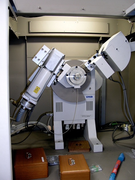
X-ray Powder Diffractometer for Multi-sample Measurement (XRD)
Mark: X
Instrument: X-ray Powder Diffractometer, Rigaku RINT-2100
Features: This is an analytical device that measures X-ray diffraction patterns of minerals, and enables following analyses.
(1) Identifies materials by comparing known X-ray diffraction pattern
(2) Differentiates materials with the same chemical composition but different structure
(3) Analyses constituent phase
(4) Identifies structure of unknown substance by combining Rietveld Method and Direct Method
This system possesses an X-ray powder database of natural and synthesized material (ICDD PDF-2, 2023), and enables computer searching from X-ray diffraction patterns obtained from experiments.
Related classes: Practical: Earth’s Environmental Chemistry (Undergraduate), Laboratory Experiments for Instrumental Analysis I/II (Graduate)
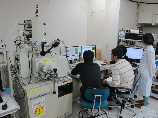
Electron Probe Micro Analyzer (FE-EPMA)
Mark: E
Equipment: Electron Probe Micro Analyzer (FE-EPMA), JEOL JXA-8530F
Features: This system is equipped with a field emission (FE) electron gun, which enables analysis in the sub-micrometer range. This instrument has the following detectors arranging around the sample chamber, and various information can be obtained. (1) Secondary electron detector (2) Backscattered electron (BSE) detector (3) 5-channel Wavelength dispersive X-ray (WDS) detector (1ch LDE2H/TAPH, 2ch LDE1/TAP, 3ch PET/LIF, 4ch PET/LIF, 5ch PETH/LIFH) (4) Energy dispersive X-ray (EDS) detector (4) Cathodoluminescence (CL) detector (Panchromatic/3-color filter can be attached) (5) Cathodoluminescence spectroscopy
Related Courses: Laboratory Experiments for Instrumental Analysis I/II (Graduate), Instrumental analyses of solids (Undergraduate/Graduate)
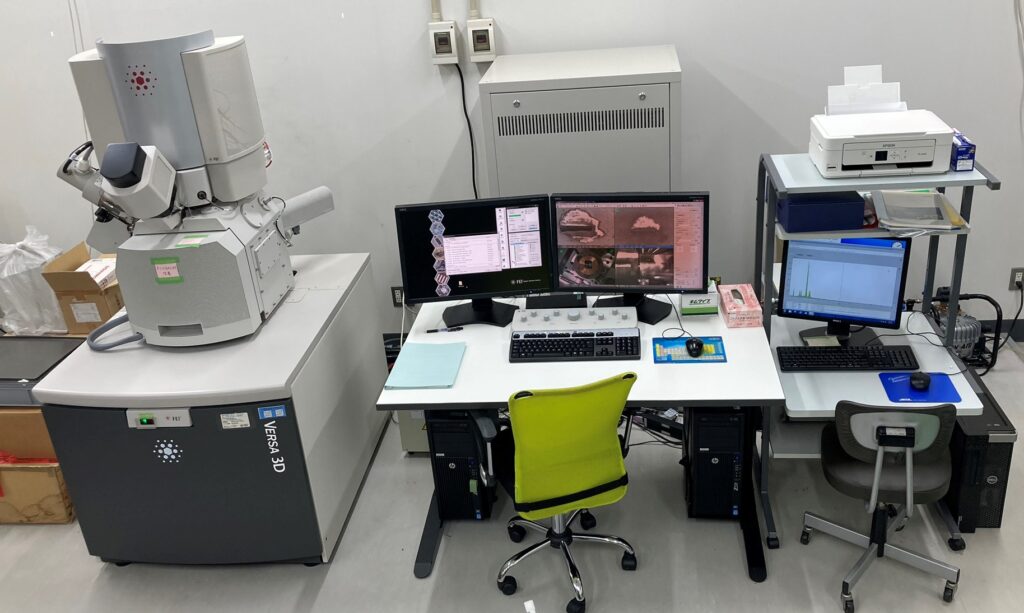
Dual beam for processing and observation (FIB-SEM)
Mark: F
Equipment: Thermo Fisher Scientific K.K., Versa 3D DualBeam
Features: In addition to the scanning electron microscope (SEM) function equipped with a field emission electron gun, focused ion beam processing (FIB) with a Ga ion gun and manipulator work with a probe are available. This instrument has a large sample chamber and can insert a sample several centimeters in height. The following detectors are placed around the sample chamber to obtain various information from the sample: (1) secondary electron detector (ET detector) (2) concentric backscatter (CBS) detector (3) energy dispersive X-ray (EDS) detector.
Related Courses: instrumental analysis practice, earth and planetary environmental science and practice
Scanning Transmission Electron Microscopy (TEM/STEM)
Mark: T
Instrument: JEM-2800 High Throughput Electron Microscope
Features: JEM-2800 is an electron microscope equipped with a Schottky-emission electron gun (200 keV), which has both Scanning Transmission Electron Microscope_(STEM) and Transmission Electron Microscope_(TEM) modes. In addition, energy dispersive X-ray spectroscopy_(EDS)with 100 mm2 dual silicon drift detectors (SDD).
(SSD) is also available. Concretely, the following analyses are possible:
- Imaging of thin films or fine particles (resolution: 0.1 nm for transmission images, 0.16 nm for STEM bright-field and dark-field images, 0.05 nm for secondary electron images)
- Crystal structure analysis based on observation of electron diffraction patterns
- Fast elemental mapping and composition analysis using EDS
Related classes: practical crystallography

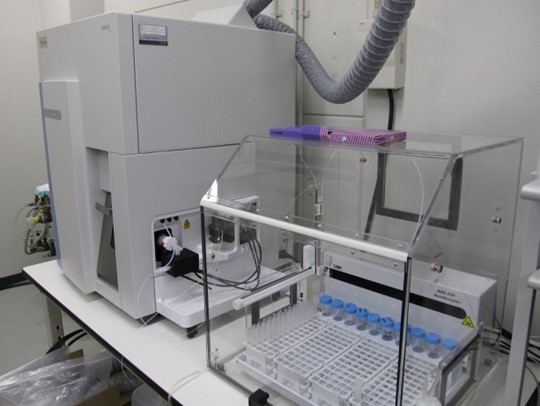
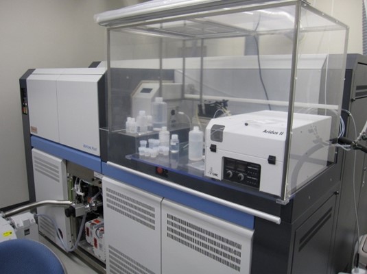
Laser Ablation-Plasma Ion Source Mass Spectrometer (LA-ICPMS)
Mark: M
Instrument: Thermo Fisher Scientific Neptune Plus and Agilent 8900
Features: ICPMS is a mass spectrometer that ionizes elements with high-temperature argon plasma and divides the ions by mass by electric or magnetic field to determine the element concentration and isotope composition. Argon plasma has a very high temperature and can efficiently ionize most elements (other than rare gasses and halogens). Furthermore, since the plasma ion source is under atmospheric pressure, this device can be combined with various sample injection methods. By combining with the laser ablation method, trace element concentrations and isotope composition of solid samples such as minerals and glass can be determined with high spatial resolution (~ 10 µm).
Related Courses: Practical: Earth and Planetary Environmental Science (Undergraduate), Laboratory Experiments for Instrumental Analysis I/II (Graduate)
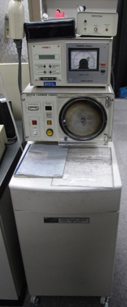
Carbon coater
Mark: P
Instrument: Quick carbon coater (Sanyu, SC-701C)
Features: Carbon is coated on the surface of observation/analysis samples for scanning electron microscope and EPMA (P).
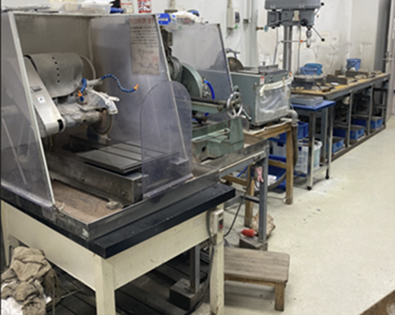
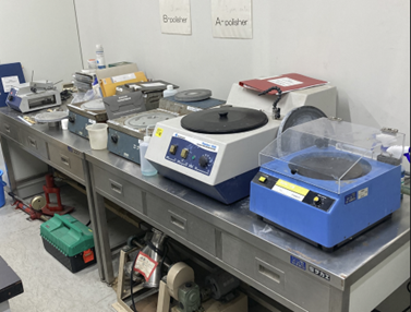
Rock processing and thin section preparation room and
Electron microscope sample preparation room
Mark: R1,R2
A large oil cutter for cutting rocks, an automatic polishing table, and a work table for manual polishing can be used to prepare thin sections from rocks collected in the field for optical microscope observation (rock processing room and student thin section production room). In addition, precision cutting of small rock fragments, resin embedding, processing in a vacuum oven, and preparation of thin sections for analysis for several electron microscope and microprobe systems (such as SEM, EPMA and FIB-SEM) can be performed (Electron microscope sample preparation room).
Related Courses: Field Excursion: Earth and Planetary Environmental Science I/II/III (Undergraduate), Field Exercise: Earth and Planetary Environmental Science I/II/III, Practical: Earth and Planetary Environmental Science (Undergraduate)